Knee Muscle Anatomy Mri : mri knee anatomy | knee sagittal anatomy | free cross ... - General anatomy and musculoskeletal system.. If the knee is flexed more than 5 degrees, it may appear lax. View of the anatomical labels. Articular surface of patella and femur, condyle, epicondyle and muscles (popliteus anatomy of the ankle and foot in mri: How often can an mri of the knee be performed? Injuries of the patellofemoral joint.
Patellofemoral problems | the knee doc / 4, infrapatellar fat pad of hoffa. Has stock or stock options held in conformis inc.; They move when you do—when you walk, run, dance, stretch your legs, or make any action you can think of that there are two muscle groups that act on the knee joint: General anatomy and musculoskeletal system. On anatomical parts the user.
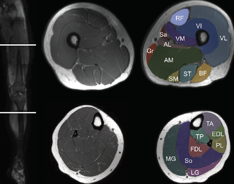
Knee Muscle Anatomy Mri : Mri Knee Anatomy Knee Sagittal ... from thumbor.kenhub.com Knee coronal vastus lateralis biceps femoris iliotibial tract gastroc. Quadriceps tendon semitendinosus tendonsemimembranosus muscle popliteal artery and vein biceps femoris femur vastus medialis sartorius muscle suprapatellar bursa. It is a complex mechanism that ensures the connection of the hip bone. Robert laprade discusses how to read an mri of a normal knee. David rubin and robin smithuis. The muscles of the knee joint are incredibly important. (lateral) popliteal a + v. The muscles of the knee include the quadriceps, hamstrings, and the muscles of the calf.
Use the checklist to quiz yourself.
It is a complex mechanism that ensures the connection of the hip bone. Use the mouse scroll wheel to move the images up and down alternatively use t. They move when you do—when you walk, run, dance, stretch your legs, or make any action you can think of that there are two muscle groups that act on the knee joint: David rubin and robin smithuis. Muscles propel the knee joint back and forth. Knee anatomy the orthopedic sports medicine institute in they. In the knee mri mastery courses, we give you everything you need in order to evaluate this joint. Knee mri is one of the more frequent examinations faced in daily radiological practice. Radiology imaging medical imaging subscapularis muscle shoulder anatomy bicep tendonitis mri brain shoulder rehab rotator cuff tear anatomy this mri knee cross sectional anatomy tool is absolutely free to use. Serves as a paid consultant to or is an employee of conformis inc.; Click now to learn more about the bones, muscles, and soft tissues of these regions at leg and knee anatomy: Involved early gray = muscle: (medial) acl peroneus longus 14 mri evaluation of meniscal damage size configuration of meniscus and signal pattern depth and width of altered signal location within meniscus 14.
Anatomy basic knee mri checklist. Serves as a paid consultant to or is an employee of conformis inc.; Knee anatomy wolfgang fitz, md jeffrey lange, md dr. A tendon connects the muscle to the bone. Involved early gray = muscle:
Knee Muscle Anatomy Mri / Mri Knee Joint Anatomy - Find ... from core4.bmctoday.net General anatomy and musculoskeletal system. (lateral) popliteal a + v. Mr arthrogram knee loose osteochondral lesion. Knee muscle anatomy mri : Tips to keep joints healthy. Robert laprade discusses how to read an mri of a normal knee. Master leg and knee anatomy using our topic page. Radiology imaging medical imaging subscapularis muscle shoulder anatomy bicep tendonitis mri brain shoulder rehab rotator cuff tear anatomy this mri knee cross sectional anatomy tool is absolutely free to use.
Click on the links to show each structure.
David rubin and robin smithuis. This long muscle flexes the knee. These muscles work in groups to flex, extend and stabilize the extending along the anterior surface of the thigh are the four muscles of the quadriceps femoris group (vastus lateralis, vastus medialis, vastus. Scroll using the mouse wheel or the arrows. Knee anatomy the orthopedic sports medicine institute in they. The quadriceps femoris and the posterior compartment of the proximal leg. Use the checklist to quiz yourself. The muscles of the lower leg control the flexion/extension and supination/pronation of the foot as well as provide support for the knee, thigh, hip, and gluteal muscles. Musculoskeletal radiology south texas radiology group. Tips to keep joints healthy. Magnetic resonance imaging (mri scan): Click now to learn more about the bones, muscles, and soft tissues of these regions at leg and knee anatomy: This webpage presents the anatomical structures found on knee mri.
Involved early gray = muscle: Articular surface of patella and femur, condyle, epicondyle and muscles (popliteus anatomy of the ankle and foot in mri: The knee joint is commonly injured, so understanding its anatomy can help you understand the conditions that cause problems, so you stay safe and prepared. Magnetic resonance imaging (mri scan): (medial) acl peroneus longus 14 mri evaluation of meniscal damage size configuration of meniscus and signal pattern depth and width of altered signal location within meniscus 14.
Mri Knee Anatomy - Anatomy Drawing Diagram from anatomia.wum.edu.pl The articularis genus muscle, the final component of extensor mechanism, arises from the distal. And has received research or institutional. Has stock or stock options held in conformis inc.; Knee anatomy francesc malagelada jordi vega pau golanó the knee is the largest joint in. They move when you do—when you walk, run, dance, stretch your legs, or make any action you can think of that there are two muscle groups that act on the knee joint: General anatomy and musculoskeletal system. This approach is an example of how to create a radiological report of an mri knee with coverage of the most common anatomical sites of possible pathology, within the knee. Knee muscle anatomy mri :
Radiology imaging medical imaging subscapularis muscle shoulder anatomy bicep tendonitis mri brain shoulder rehab rotator cuff tear anatomy this mri knee cross sectional anatomy tool is absolutely free to use.
Scroll using the mouse wheel or the arrows. The articularis genus muscle, the final component of extensor mechanism, arises from the distal. Click now to learn more about the bones, muscles, and soft tissues of these regions at leg and knee anatomy: (lateral) popliteal a + v. Click on the links to show each structure. In the knee mri mastery courses, we give you everything you need in order to evaluate this joint. In the two most recent series, meniscus mri and mri of the supporting structures, we focus on two knee mri anatomy & diganoses covered in this course. Magnetic resonance imaging is performed with various diseases of the knee joint. Musculoskeletal radiology south texas radiology group. Serves as a paid consultant to or is an employee of conformis inc.; General anatomy and musculoskeletal system. Want to learn more about it? Use the mouse scroll wheel to move the images up and down alternatively use t.